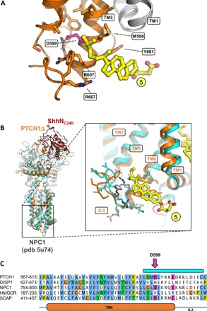
Fig. 3. The inner leaflet SSD sterol site points at a functional role in PTCH1.

(A) The site 5 sterol (as defined in Fig. 2B; colored yellow) is within contact of the residue D599, previously shown to be functionally critical in the SSDs of SCAP and Drosophila Patched. (B) A similar site, unoccupied by a sterol molecule, has been previously observed in the structure of the cholesterol transporter NPC1, homologous to PTCH1. The PTCH1Δ-ShhNC24II structure was aligned to the NPC1 X-ray structure (cyan). Inset: Alignment of the two structures using the residues of TM6 shows a great degree of similarity in the region surrounding the site 5 sterol. (C) Sequence alignment of key SSD-containing proteins, PTCH1, DISP1, NPC1, HMG-CoA reductase, and SCAP, shows the similarity between the elements involved in inner leaflet sterol binding site. The pink arrow indicates the residue D599 [shown in (A)]. The cyan bar corresponds to residues shown with side chains in (B). TM6 and IL3 of PTCH1 are indicated below the sequence alignment.
