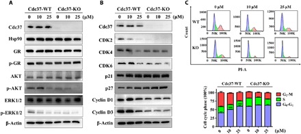
Fig. 4. DDO-5936 arrests the cell cycle in HCT116 cells.

(A) Western blot analysis of Cdc37, Hsp90, GR, p-GR, AKT, p-AKT, ERK1/2, and p-ERK1/2 protein expression levels in Cdc37-WT HCT116 cells and corresponding Cdc37-KO HCT116 cells after treatment with 0, 10, or 25 μM DDO-5936 for 24 hours. β-Actin was used as a loading control. Data are representative of three independent experiments. (B) Western blot analysis of Cdc37, CDK2, CDK4, CDK6, p21, p27, cyclin D1, and cyclin D3 protein expression levels in Cdc37-WT HCT116 cells and corresponding Cdc37-KO HCT116 cells after treatment with 0, 10, or 25 μM DDO-5936 for 24 hours. β-Actin was used as a loading control. Data are representative of three independent experiments. (C) Top: Cell cycle distribution measured by propidium iodide (PI) staining of Cdc37-WT HCT116 cells and corresponding Cdc37-KO cells after treatment with 0, 10, or 25 μM DDO-5936 for 24 hours. Bottom: The cell cycle distribution is represented as a graphic histogram. Data are representative of three independent experiments as the means ± SD (***P < 0.001).
