. 2019 Sep 5;8:e50231. doi: 10.7554/eLife.50231

Figure 2. Inhibiting macrophage movement with latrunculin A or blebbistatin abolishes particle release onto the surrounding substrate.

Scanning electron micrographs (SEMs) of mouse peritoneal macrophages that had been treated with latrunculin A (A) or blebbistatin (B). Macrophages were treated with latrunculin A, blebbistatin, or vehicle alone (DMSO) overnight; the drug treatments were initiated when the cells were in suspension 1 h before plating ('pre-treatment') or 1 h after adherence to the substrate ('post-adherence'). Macrophages treated with vehicle alone (DMSO) released particles onto the substrate. No particles were visible on the substrate in cells that had been pre-treated with latrunculin A or blebbistatin—or in the blebbistatin post-adherence cells. In the latrunculin A post-adherence cells, a circumferential ring of particles was observed around cells. Latrunculin A and blebbistatin were removed from some dishes after the overnight incubation, and the cells were incubated for an additional 18 h without drugs. After removing the drugs, the release of particles onto the substrate resumed. Four independent experiments were performed; representative images are shown. Scale bars for images on the left in each panel are 4 µm; scale bars for the images in the middle and right are 1 µm.

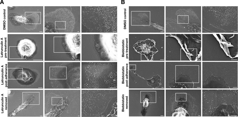
Figure 2.

Figure 2—video 1. Treatment of mouse peritoneal macrophages with latrunculin A or blebbistatin prevent projection and retraction of filopodia/lamellipodia.
Download video file (1.3MB, mp4)
DOI: 10.7554/eLife.50231.007
Video shows macrophages treated with vehicle (DMSO) alone. Macrophages were plated on glass-bottom Petri dishes and incubated in medium containing latrunculin A (5 μM), blebbistatin (30 μM), or vehicle (DMSO) alone. Cells were imaged by live-cell microscopy for 18 h at 5-min intervals. Movement of macrophage lamellipodia was observed in the 'DMSO Control' macrophages but not in macrophages treated with latrunculin A or blebbistatin. Videos show an 18-h period of live-cell imaging.
Video shows macrophages treated with vehicle (DMSO) alone.
Figure 2—video 2. Shows macrophages treated with vehicle (DMSO) alone.
Download video file (2.2MB, mp4)
DOI: 10.7554/eLife.50231.008
Figure 2—video 3. Shows macrophages treated with latrunculin A.
Download video file (4.6MB, mp4)
DOI: 10.7554/eLife.50231.009
Figure 2—video 4. Shows macrophages treated with latrunculin A.
Download video file (4.9MB, mp4)
DOI: 10.7554/eLife.50231.010
Figure 2—video 5. Shows a macrophage treated with blebbistatin.
Download video file (2.6MB, mp4)
DOI: 10.7554/eLife.50231.011
Figure 2—video 6. Shows macrophages treated with blebbistatin.
Download video file (4MB, mp4)
DOI: 10.7554/eLife.50231.012