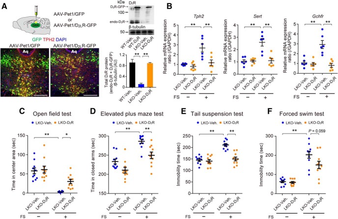
Figure 6.
Overexpression of D2LR in 5-HT neurons of the DRN reduces stress vulnerability in D2LR-KO mice. A, Left, Representative immunofluorescence images showing AAV-Pet1/GFP (left) and AAV-Pet1/D2LR-GFP (right) expression in the DRN. Green, GFP; red, TPH2; blue, DAPI; Aq, aqueduct. Scale bars, 250 μm. Right, Top, Representative immunoblot of mouse DRN lysates probed with the indicated antibodies. Bottom, Densitometric analysis of D2R levels normalized to β-tubulin (A.U., arbitrary units). **p < 0.01 by one-way ANOVA with Bonferroni's post hoc test. n = 3 mice each. endo-D2R, Endogenous D2R. B, RT-qPCR analysis of mRNA levels in mouse DRN lysates. n = 6 mice each. Each bar represents the mean ± SEM. **p < 0.01 by two-way ANOVA with Bonferroni's post hoc test. C–F, Local expression of D2LR in the DRN ameliorates stress vulnerability in D2LR-KO mice in the open-field test (C), elevated plus maze test (D), tail-suspension test (E), and FS test (F). n = 10 mice each. Each bar represents the mean ± SEM. **p < 0.01, *p < 0.05 by two-way ANOVA with Bonferroni's post hoc test.

