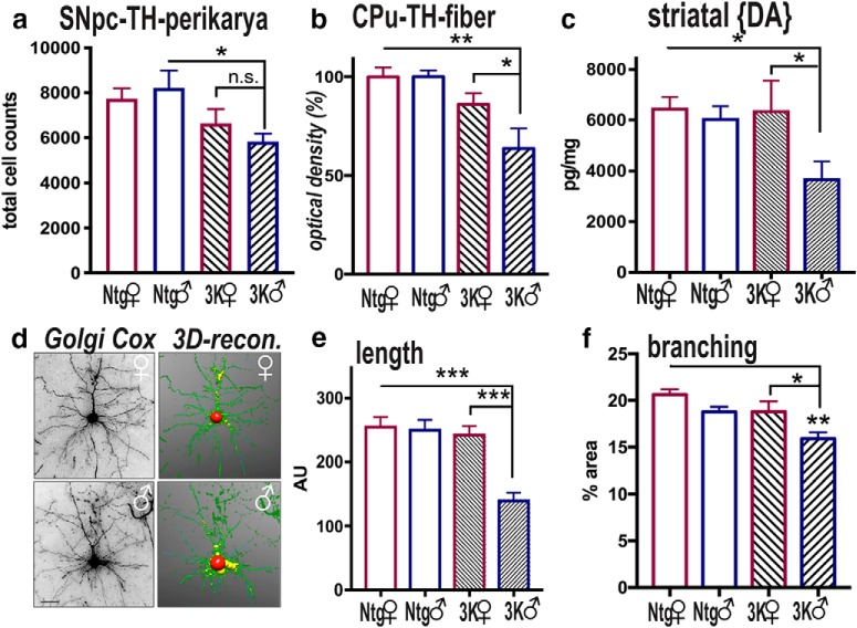
Figure 3.
Sex differences in cortical and dopaminergic neurodegeneration in 6-month-old 3K mice. a, Unbiased stereological counting determined the number of TH+ neurons in the SNpc of 6-month-old mice. b, Relative TH optical density (total of 18 sections each group) was analyzed in the CPu. c, HPLC assay of striatal dopamine in 6-month-old male and female 3K mice vs. Ntg littermates (a–c: Ntg, 3K n = 5 each sex). d, Using NeuronStudio software for automatic tracing of Golgi Cox-impregnated neuritic profiles for (e) estimating fiber length (n = 10 traced neurons) or (f) percentage area covered (n = 10 fields) of cortical sections (n = 3 mice per age and genotype). Data are mean ± SEM. Two-way ANOVA, post Tukey: n.s., non-significant; *p < 0.05; **p < 0.01; ***p < 0.001. Scale bar: d, 25 μm.

