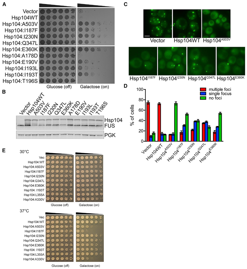
Figure 3. Hsp104 NBD1 Variants Suppress FUS Toxicity and Aggregation.
(A) NBD1 variants suppress FUS toxicity in yeast. W303aΔhsp104-pAG-303GAL-FUS yeast were transformed with Hsp104 variants or vector. Strains were serially diluted 5-fold and spotted in duplicate onto glucose (non-inducing) and galactose (inducing) media.
(B) NBD1 variants do not grossly reduce FUS expression in yeast. Strains in (A) were induced for 5 h, lysed, and immunoblotted for Hsp104, FUS, and PGK (loading control).
(C) NBD1 variants suppress FUS aggregation in yeast. Fluorescence microscopy of cells coexpressing FUS-GFP and Hsp104 variants. Strains were induced for 5 h in galactose and imaged. Scale bar, 2.5 μm.
(D) FUS aggregation was quantified by calculating the proportion of cells containing multiple foci, a single focus, or no foci. Values represent means ± SEM (n = 2–3).
(E) Potentiated NBD1 variants typically do not exhibit reduced growth at 37°C. Hsp104 variants were expressed in the 416GAL vector in Δhsp104 yeast in the absence of any disease protein. Strains were serially diluted 5-fold and spotted in duplicate onto glucose (non-inducing) and galactose (inducing) media and grown at 30°C or 37°C.
See also Figures S1 and S2.

