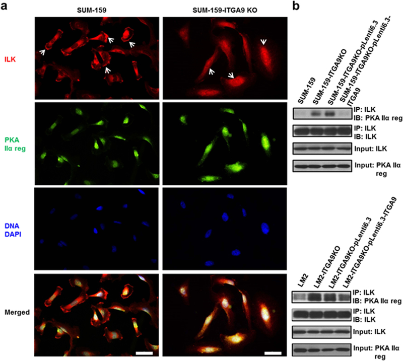
Fig. 6.

ITGA9 KO causes ILK relocation from cellular membrane region to cytoplasm and increases the interaction between ILK and PKA regulatory subunit IIα. a Representative images of IF staining of ILK (red), PKA regulatory subunit IIα (green) and nuclear DNA DAPI (blue) in parental and ITGA9 KO SUM-159 cells. White arrows point to representative ILK cellular membrane staining in parental cells and ILK cytoplasm staining in ITGA9 KO cells. Scale bar, 50 μm. b Representative Western blot images of Co-IP analysis determining the interaction between ILK and PKA regulatory subunit IIα in parental, ITGA9 KO and re-expressing LM2 and SUM-159 cells. IP: immunoprecipitating; IB: immunoblotting.
