Figure 2.
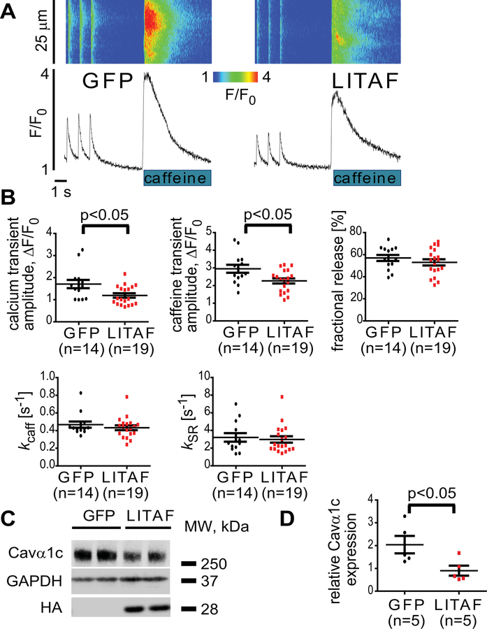
Attenuation of Ca2+ transients and Cavα1c abundance by LITAF in adult rabbit cardiomyocytes. Cardiomyocytes were transduced with adenovirus expressing GFP or HA-LITAF. A, Representative confocal line scan images and corresponding Fluo-3 F/F0 time-dependent profiles at 1 Hz. B, Histograms depict mean data from Ca2+ transient amplitudes (GFP, 1.78±0.16 vs LITAF, 1.2±0.13 ΔF/F0), caffeine transient amplitudes (GFP, 2.8±0.15 vs LITAF, 2.1±0.19 ΔF/F0), fractional release and rates of Ca2+ removal by NCX (kcaff) and SERCA2 (kSR). Student’s t-test, p<0.05 (2–3 heart preparations). C, ARbCM lysates were probed with anti-Cavα1c, anti-HA and anti-GAPDH to indicate Cavα1c, exogenous LITAF and GAPDH (loading control) protein levels. D, Respective change in Cavα1c abundance, normalized to GAPDH (n=5 animals, performed in triplicate; mean±SEM). Student’s t-test, p<0.05.

