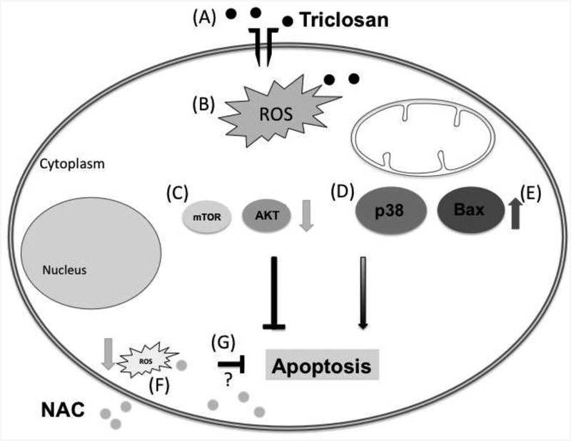
Fig. 6. Signaling pathways altered by TCS exposure.
Upon exposure to 50 μM TCS for 12 h, PC12 cells viability was decreased (A), and ROS production was increased (B). Both antiapoptotic proteins mTOR and Akt (C) display decreased phosphorylation. We also observed increased phosphorylation of p38 MAPK (D), which has been shown to participate in apoptosis. Due to increased expression of Bax (E), it is likely that apoptosis takes place. Treatment with N-acetyl cysteine (NAC) decreased ROS in this model (F). However it remains to be determined whether oxidative stress plays a role in the alterations observed in this study (G).

