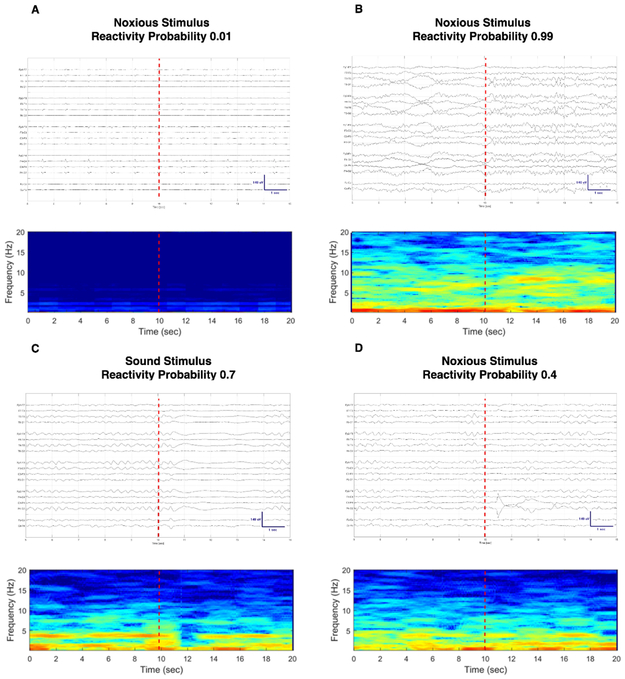
Figure 4:
Compressed spectral array display and raw EEG signal with corresponding probability of good outcome using QEEG reactivity as a risk score. 4A: subject had poor outcome and a low neurological recovery score; 4B: subject with good outcome and high neurological recovery score; 4C and 4D: subject with good outcome who had a higher neurological recovery score for sound vs. pain stimuli. There is a diffuse decrease in amplitude for three seconds after sound stimulation (4C). No definite change on EEG is observable after noxious stimulation by visual inspection of the EEG recording (4D).

