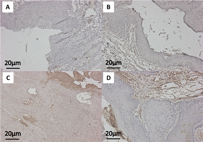
Figure 3: Represenatative immunohistochemistry images of pS6 expression.
Images show anal tissue from K14E6/E7 mice stained for pS6 (brown) protein expression with hematoxylin counterstaining (blue). The panels correspond with the following groups: A) No treatment (Control), B) Topical BEZ235, C) DMBA, and D) DMBA+Topical BEZ235.

