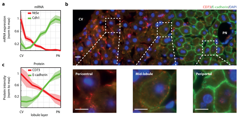
Fig. 2. CD73 and E-cadherin are inversely zonated surface markers.
a, CD73, encoded by Nt5e, and E-cadherin, encoded by Cdh1, are surface markers that are zonated at the mRNA level. Data taken from 5, n=1415 cells from 3 mice. Lines show sum-normalized mean of all cells, shaded regions are ±SEM. b, CD73 and E-cadherin proteins are zonated. Shown is an example of a lobule stained by immunofluorescence with antibodies against CD73 (red) and E-cadherin (green). Blue – DAPI nuclear stain. Scale bar – 10μm. Experiment was prformed independently on 3 different mice. c, Quantification of immunofluorescence images (n=8 lobules from three mice). Lines represent the mean of intensity measured in the lobule layer, shaded regions are ±SEM across the eight lobules. CV – central vein, PN – portal node.

