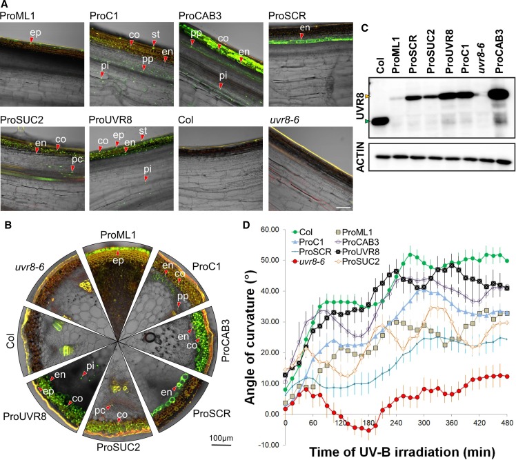
Figure 6.
Stem Bending Phenotype of the uvr8 Mutant Can Be Complemented in YFP-UVR8 Expression in Different Cell Types.
(A) Longitudinal sections made from Arabidopsis inflorescence stems demonstrate the expression pattern of YFP-UVR8 expressed under the control of different promoters in the uvr8-6 background. Col and uvr8-6 are presented as controls. The presented images are the overlay of the green and bright-field channels obtained from CLSM. Green color indicates the YFP signal; red color shows autofluorescence. A few representative nuclei containing YFP-UVR8 are marked in the epidermis (ep), stomata (st), cortex (co), endodermis (en), phloem parenchyma (pp), phloem companion cells (pc), and pith (pi). Bar = 100 µm.
(B) Transverse sections of the same stems as depicted in (A). The marked nuclei in different tissues are named also as in (A).
(C) Immunoblot analysis of UVR8 and YFP-UVR8 expression levels in the inflorescence stems. (Top) Result of the membrane hybridization using the anti-UVR8 antibody, with the green arrowhead indicating native UVR8 and the orange arrowhead indicating YFP-UVR8. (Bottom) Result of immune staining using anti-ACTIN antibody, demonstrating the equal total protein amounts in the lanes.
(D) Kinetic analysis of the bending response of Arabidopsis inflorescence stems exposed to unilateral UV-B treatment. Inflorescence stem reorientation was quantified over time. Error bars show se (n ≥ 8). The full names of the transgenic lines throughout this figure are as follows: ProML1, ProML1:YFP-UVR8/uvr8-6; ProC1, ProC1:YFP-UVR8/uvr8-6; ProCAB3, ProCAB3:YFP-UVR8/uvr8-6; ProSCR, ProSCR:YFP-UVR8/uvr8-6; ProUVR8, ProUVR8:YFP-UVR8/uvr8-6; ProSUC2, ProSUC2:YFP-UVR8/uvr8-6.

