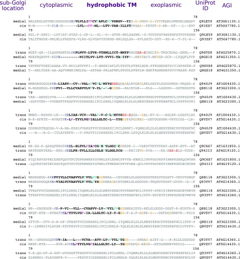
Figure 6.
Comparison of Type II TM Protein Paralogues with Different Sub-Golgi Classifications.
Alignments are shown for pairs of similar, homologous proteins from Arabidopsis that have different sub-Golgi localizations. TM span regions are indicated in boldface. The blue Arg/Lys at the cytoplasmic edge highlight the start of the TM span. Phe residues are colored either pink or cyan to indicate relative position in the TM span. Within 15 residues of the exoplasmic TM edge, Ser residues are colored yellow and three consecutive Ser residues are colored red.

