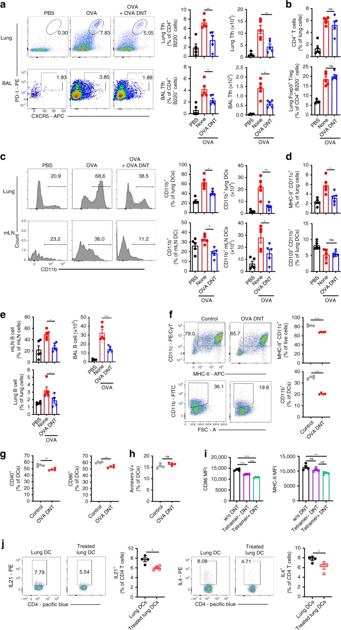
Fig. 4.
OVA DNT treatment selectively inhibited Tfh cells and CD11b+ DCs. OVA-sensitized mice were treated with an intravenous transfer of OVA DNTs after the first OVA challenge. The mice were challenged daily for the next two days and sacrificed 48 h after the last aerosol challenge. a The lung and BALF Tfh cell (CD4+B220-CXCR5+PD-1+), b CD4+ T cell (CD4+B220-) and Treg cell (CD4+B220-Foxp3+) proportions were measured by flow cytometry. c The lung and mLN CD11b+ DC (CD11c+MHC-II+CD11b+), d DC (CD11c+MHC-9II+) and CD103+ DC (CD11c+MHC-II+CD103+CD11b-) proportions were measured by flow cytometry. e The proportions of B cells (B220+CD4-) in the mLN (mediastinum lymph node), BALF and lungs were measured by flow cytometry. OVA-stimulated bone marrow cells were cocultured with OVA DNTs and stimulated with GM-CSF (20 ng/ml) for 3 days to test the direct effect of OVA DNTs on OVA DC proliferation and differentiation. f The proportions of bone marrow-derived DCs and CD11b+ DCs were measured by flow cytometry. The direct effects of OVA DNTs on g costimulatory molecule expression and h apoptosis in DCs were measured by flow cytometry. i OVA tetramer+ and tetramer- DNT cells were sorted by flow cytometry from OVA-primed DNT cells. Tetramer+ or tetramer− DNT cells were cocultured with lung DCs from allergic asthma mice for 3 days. The MFIs of CD86 and MHC-II in DCs were measured by flow cytometry. j Lung DCs (2.5 × 10⁴) from OVA DNT cell-treated or -untreated asthma mice were cocultured with 1 × 10⁵ naive CD4+ T cells for 3 days. IL21- and IL4-secreting CD4+ T cells were measured by flow cytometry. Data are shown as the mean ± SEM; n = 4–5 mice per group. One-way ANOVA and Student’s t-test were used to calculate significance. *P < 0.05; **P < 0.01; ***P < 0.001. Source data are provided as a source data file

