Fig. 6.
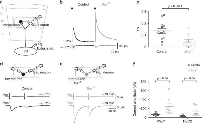
An exceeding number of interneurons increases inhibition in neuronal networks. a Diagram representing the experimental procedure where a layer IV glutamatergic neuron recorded during the extracellular stimulation of the ventro-basal nucleus (VB) of the thalamus. In this circuit, the recorded neuron is synaptically connected by FSI which also receive thalamic input that triggers a strong disynaptic feedforward inhibition. b Excitatory (inward) and inhibitory (outward) PSCs evoked by thalamic stimulation in layer IV glutamatergic neurons of control (black) and Baxf/f mice (dark gray) recorded at −70 and 0 mV, respectively, in acute thalamocortical slices during the third postnatal week. Stimulation artifacts were blanked for visibility. The stimulation time is indicated (arrowheads). c Dot plots of excitation/inhibition (E/I) ratio obtained for glutamatergic neurons in control (black, n = 13) and Baxf/f (dark gray, n = 10) mice by dividing excitatory PSCs by inhibitory PSCs (Mann–Whitney U test; significant p-value is indicated). d, e Paired recordings between a presynaptic FSI and a layer IV glutamatergic neuron in control (black, d) and Baxf/f (dark gray, e) mice. Note that action currents evoked in FSI elicited larger PSCs in glutatamergic neurons of Baxf/f mice. f Dot plots of PSCs evoked by the first (PSC1) and second (PSC2) action current in the FSI (Mann–Whitney U test; significant p-value are indicated). The paired-pulse ratio (PPR = PSC1/PSC2) was not different between control (black, n = 4 out of 8 pairs connected) and Baxf/f (dark gray, n = 7 out of 7 pairs connected) mice, indicating that there were no changes in the release probability of presynaptic FSI (PPR: 0.45 ± 0.05 and 0.52 ± 0.07, respectively; p = 0.412, Mann–Whitney U test). Data are presented as mean ± SEM

