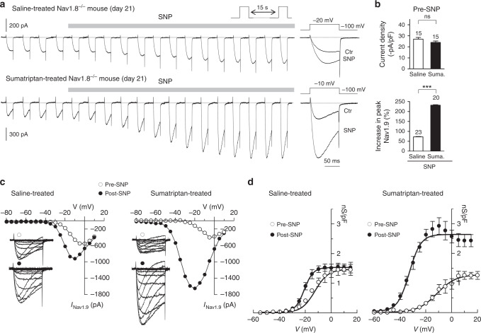
Fig. 4.
Sumatriptan treatment promotes activation of Nav1.9 by NO. a Nav1.9 current exposed to 1 mM SNP in dural afferent neurons from saline-treated (top panel) and sumatriptan-treated (bottom panel) Nav1.8−/− mice. CsCl-only-based patch pipette solution. Right-most traces: superimposed Nav1.9 currents before and after SNP application. b Nav1.9 current density (top panel) in dural afferent neurons from saline-treated and sumatriptan-treated Nav1.8−/− mice. ns, not significant; unpaired t-test. Bottom panel: mean increase in Nav1.9 peak current induced by SNP (1 mM) in dural afferent neurons from saline-treated and sumatriptan-treated Nav1.8−/− mice. ***p < 0.001; unpaired t-test. c Nav1.9 I–V determined in dural afferent neurons from saline-treated and sumatriptan-treated Nav1.8−/− mice before and after SNP exposure. Insets: superimposed Nav1.9 current traces evoked by voltage steps from −80 to +10 mV from a Vh of −100 mV. Note that not all traces are shown for clarity sake. d Activation curves for Nav1.9 current determined in DiI+-dural neurons before and after SNP application. Boltzmann fits gave V1/2 values of −14.47 ± 1.3 and −20.8 ± 0.8 mV before and after SNP in saline-treated Nav1.8−/− mice (n = 11) and of −11.3 ± 1.8 and −32.7 ± 1.02 mV before and after SNP in sumaptriptan-treated Nav1.8−/− mice (n = 9), respectively. All data collected from neurons cultured at day 21

