Fig. 5.
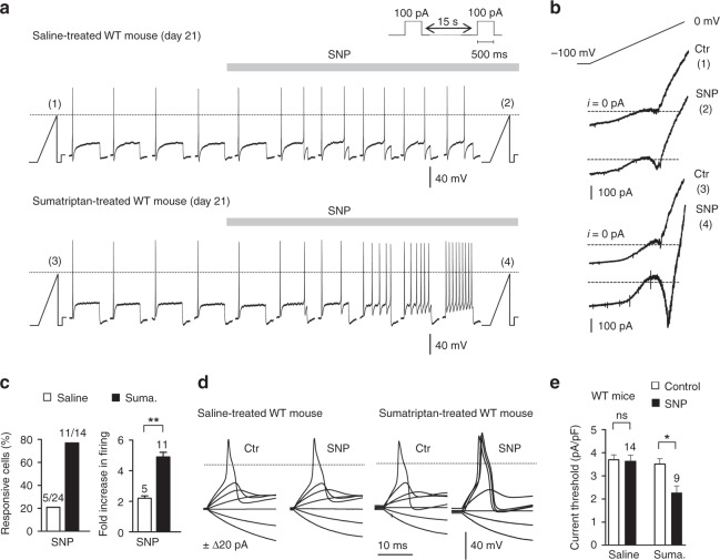
Nav1.9 activation by NO lowers excitability threshold and enhances firing in dural afferent neurons. a Effect of SNP (1 mM) on DiI+ dural afferent neurons from saline-treated (upper panel) and sumatriptan-treated (bottom panel) WT mice. KCl-based intracellular solution throughout. b I–V relationships determined using a slow (50 mV/s) voltage ramp command in DiI+ dural neurons illustrated in a before (1,3) and during (2,4) SNP application. Note the activation of Nav1.9 (inwardly flowing current) by SNP (4). c Percentage of DiI+ neurons responding to SNP (left panel) and mean change in their firing rate (right panel). Protocol as in a. **p < 0.01; Mann–Whitney test. d Generation of APs before and after SNP exposure in dural afferent neurons from saline-treated or sumatriptan-treated WT mice. Steady bias currents were used to maintain the neurons at ~−65 mV. e Comparison of normalized current threshold for AP before and during SNP application in DiI+ dural neurons from saline-treated and sumatriptan-treated WT mice. ns not significant, *p < 0.05 compared to saline-treated WT mice with Wilcoxon matched paired test. All data collected from TG neurons cultured at day 21

