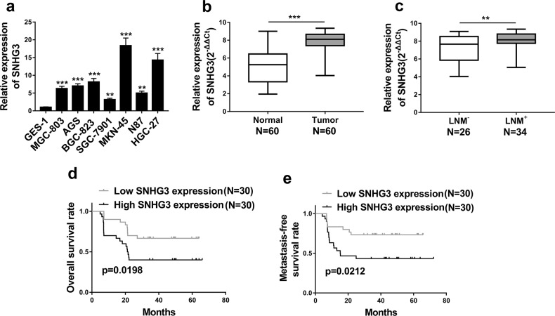
Fig. 1. SNHG3 was highly expressed in GC and increased SNHG3 level was positively correlated with poor prognosis.
a Expression of SNHG3 in various gastric cancer cell lines (MGC-803, AGS, BGC-823, SGC-7901, MKN-45, N87, HGC-27) compared to normal cell line GES-1 was detected by qRT-PCR. **P < 0.01; ***P < 0.001. b qRT-PCR assay was implemented to detect SNHG3 expression levels in 60 gastric cancer (GC) tissues (Tumor) and 60 non-tumor tissues (Normal), ***P < 0.001. c The SNHG3 expression levels were much higher in gastric cancer (GC) tissues of lymph node metastasis patients (LNM+) than patients without lymph node metastasis (LNM–) quantified by qRT-PCR analysis. **P < 0.01. d, e Kaplan–Meier survival curves demonstrated that high levels of SNHG3 were associated with poor overall survival rate and metastasis-free survival rate. *P < 0.05. Data are presented as mean ± SD and analyzed using independent samples t-test

