Figure 2.
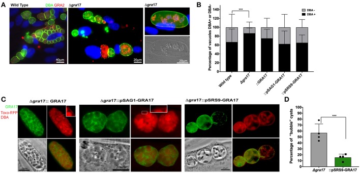
In vitro cyst formation of ME49Δgra17 and complemented strainsConfluent HFF were infected with indicated strains at a MOI of 0.2. Cyst conversions was stimulated using ambient CO2 and pH 8.1 for 5 days. (A) Samples were fixed and permeabilized with methanol, the cyst wall was stained with DBA-FITC, GRA17 was detected with a c-Myc antibody, and parasites were stained with a GRA2 antibody. Inset) higher magnification showing DBA-positive cyst wall. (B) At least 100 vacuoles for each experiment were classified as DBA-positives or DBA-negative. Each bar represents the average plus SD of biological replicates (n = 4). Significant differences in the conversion ratio between wild-type and the different strains was assessed by Student's t-test (p = 0.00012). (C) Samples were fixed with formaldehyde, the cyst wall was stained with DBA-TRITC, GRA17 was detected with a c-Myc antibody, and parasites expressed RFP. Inset) higher magnification showing DBA-positive cyst wall. (D) At least 100 cysts for each experiment were classified as regular or “bubble” cysts. Each bar represents the average plus SD of biological replicates (n = 4). Significant differences in the number of “bubble” cysts between Δgra17 and Δgra17::pSRS9-GRA17 was assessed by Student's t-test (p = 0.002).

