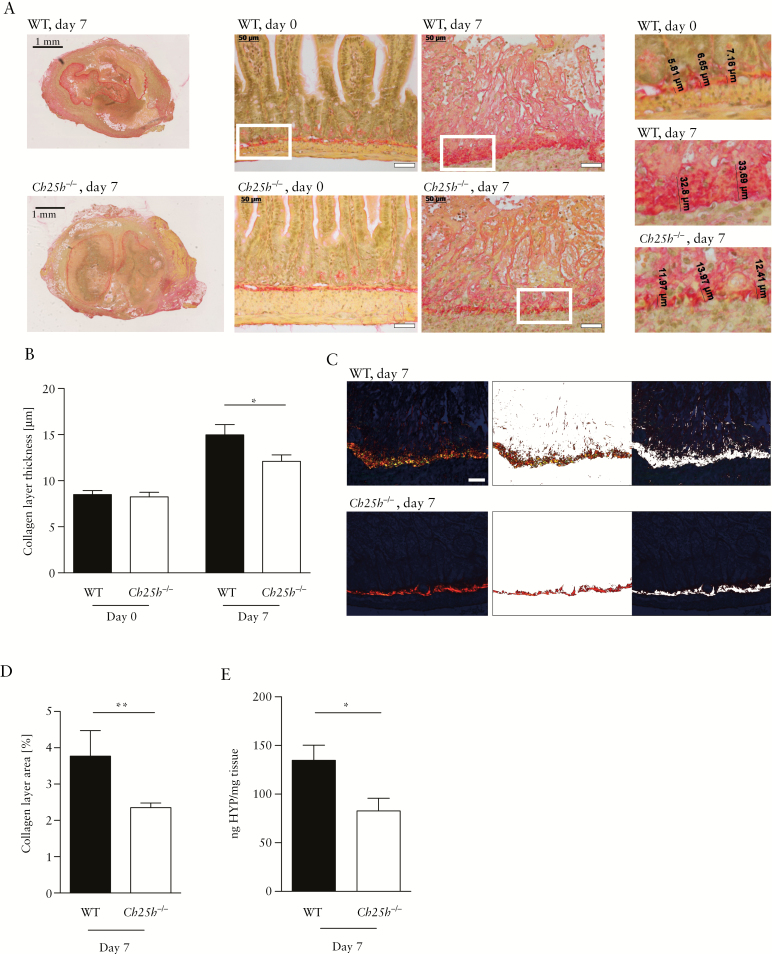
Figure 4.
Reduced levels of intestinal fibrosis in CH25H-deficient mice in the heterotopic transplantation model. Wild-type and Ch25h-/- animals were tested in a heterotopic transplantation model for intestinal fibrosis. [A] Left panels: Overview [low-resolution image] of Sirius Red-stained intestinal grafts of WT and Ch25h-/- mice at Day 7 after transplantation. Scale bar: 1 mm. Middle panels: Representative transmission light images demonstrating increased collagen layer thickness in grafts at Day 7 compared with freshly isolated intestines at Day 0. Upper panels: WT littermate controls. Lower panels: Ch25h-/-. Scale bar: 50 μm. Right panels: High-resolution inserts illustrating measurements of collagen layer thickness. [B] Collagen layer thickness calculated from eight or more positions per graft in representative areas of Sirius Red-stained slides with transmission light at 200-fold magnification. [C] Image analysis for identification of collagen layer areas using Matlab custom-made scripts. Left panel: Original polarised 200x light microscopy image. Middle panel: Collagen layer area. Right panel: Remaining non-collagen tissue. Scale bar: 50 μm. [D] Quantification of collagen layer area at Day 7 post transplantation, using the same strategy as in [C]. [E] Collagen quantification with hydroxyproline assay. Day 0, freshly isolated intestine. Day 7, intestine 7 days post transplantation [nWT day 0 = 3, nKO day 0 = 9, nWT day 7 = 8, nWT day 7 = 11]. Statistical analysis: Mann-Whitney U test; *p <0.05, **p <0.01. Bars indicate mean ± standard error of the mean [SEM]. WT, wild type; CH25H, cholesterol 25 hydroxylase; HYP, hydroxyproline.

