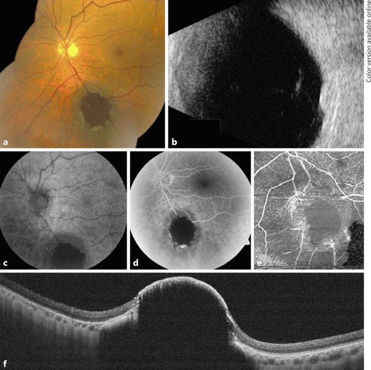
Fig. 1.
A 71-year-old male with choroidal nevus demonstrating retinal invasion. Case 1: a The pigmented choroidal tumor showed marked dark central pigmentation within the retina, hiding the retinal vessels and surrounded by trace exudation. b B-scan ultrasonography documented a dome-shaped, dense choroidal mass with a thickness of 2.7 mm. c Autofluorescence depicted central hypoautofluorescence at the site of the invasion. Fluorescein angiography (d) and optical coherence tomography angiography (e) showed central blocked perfusion corresponding to the area of invasion. f Enhanced depth imaging-optical coherence tomography through the nevus showed central invasion of the retina.

