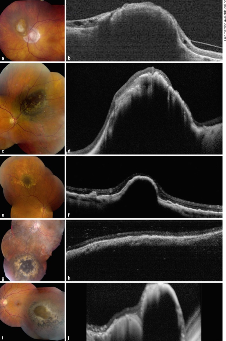
Fig. 2.
Optical coherence tomography (OCT) of choroidal nevus with retinal invasion. Case 2: clinical appearance (a) and OCT (b) of a juxtapapillary choroidal nevus with overlying fibrous metaplasia of the retinal pigment epithelium (RPE) demonstrating overlying retinal invasion and thinning. These features remained stable at 95 months' follow-up. Case 3: clinical appearance (c) and OCT (d) of a superonasal choroidal nevus with overlying patchy fibrous metaplasia of the RPE showing dark central invasion, confirmed on OCT with extreme retinal thinning and disorganization. Despite a limited follow-up period, these findings were stable 3 months later. Case 4: clinical appearance (e) and OCT (f) of a superior choroidal nevus surrounded by drusen and capped by a dark appearance representing invasion with thinning of the RPE and retina. These findings were stable at 30 months' follow-up. Case 5: clinical appearance (g) and OCT (h) of an inferior choroidal nevus with overlying fibrous metaplasia of the RPE. These findings were stable at 18 months' follow-up. Case 6: clinical appearance (i) and OCT (j) of an inferotemporal choroidal nevus with a central break of Bruch membrane and retinal invasion surrounded by RPE alterations.

