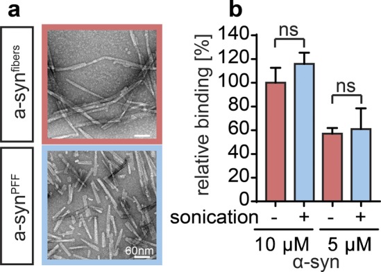
Figure S2. Characterization of sonicated α-synuclein fibrils.
(A) TEM structure of mature α-synuclein fibrils before (α-synfibers) and after sonication (α-synPFF). (B) Binding of 50 μM polyP300-AF647 to either 5 or 10 μM α-synfibers (red) or α-synPFF (blue) was measured using FP (n = 3). Relative binding of polyP to 10 μM α-synfibers was set to 100%. Comparisons were not significant by two-tailed t test.
Source data are available for this figure.

