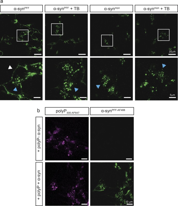
Figure S4. Localization analysis of α-synPFF and polyP by fluorescence microscopy.
(A) Differentiated SH-SY5Y cells were incubated with either 3 μM α-synPFF-AF488 or freshly purified α-synmon-AF488 for 3 h. Then, the cells were either directly imaged or after a 15-s incubation with the membrane impermeable dye trypan blue (TB), which quenches extracellular fluorescence. Brightness and contrast adjustments have been equally applied to all images. (B) Differentiated SH-SY5Y cells were incubated with or without 250 μM polyP300-AF647 for 24 h, washed, and subsequently left untreated or incubated with 3 μM α-synPFF-AF488 for another 24 h before images were taken. Brightness and contrast adjustments have been equally applied to all images.

