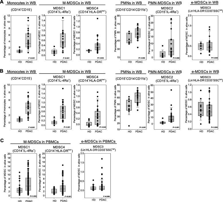
Fig. 2.
Blood-circulating MDSC enumeration in PDAC patients. a-b Flow cytometry analysis of circulating myeloid cells in whole blood of two independent cohorts of PDAC patients (b PDAC n = 21, HD = 8; c PDAC n = 23, HD = 9): monocytes (CD14+CD15−), MDSC1 (CD14+IL-4Rα+), MDSC4 (CD14+HLA-DRlow/−), granulocytes (CD15+CD14−), MDSC2 (CD15+IL-4Rα+) and MDSC3 (LIN−HLA-DR−CD33+SSChigh). c Flow cytometry analysis of circulating M-MDSCs (MDSC1, CD14+IL-4Rα+; MDSC4, CD14+HLA-DRlow/−) and e-MDSCs (MDSC3, LIN−HLA-DR−CD33+SSChigh) in PDAC patients (n = 73) compared to healthy donors (HD; n = 28). M-MDSC percentages were evaluated on frozen PBMCs, whereas e-MDSCs on the whole blood. Statistical analysis was performed by ANOVA test

