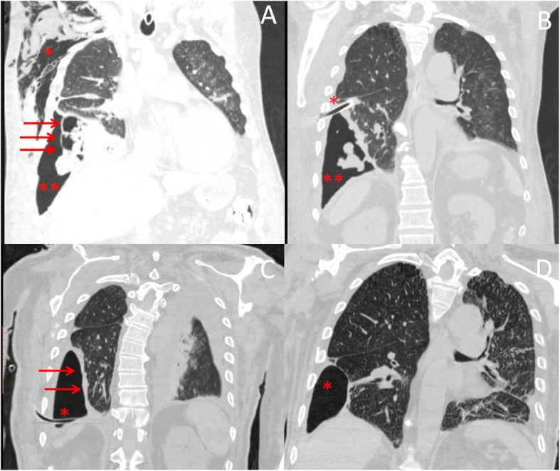
Fig. 1.

Chest computed tomography scan showed the presence of subcutaneous emphysema (*), pneumothorax (**), and necrotizing pneumonia with empyema (arrows) (Part a). After chest drainage placement (*), computed tomography scan showed the persistence of loculated pneumothorax (**) (Part b). Despite the insertion of chest tube (*), right lower lobe did not expand as it was trapped by pleural adhesions (arrows) (Part c). Following closure of alveolar pleura fistula, chest computed tomography showed no progression of loculated pneumothorax (*) (Part d)
