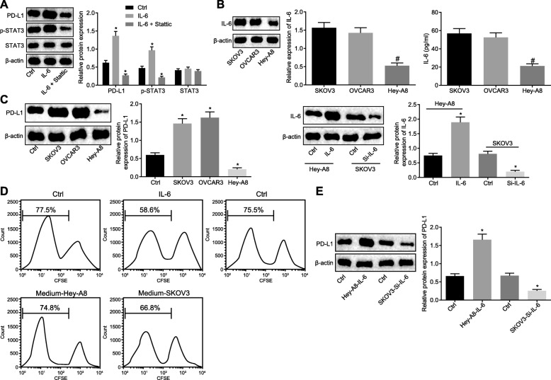
Fig. 2.
IL-6 secreted by OC cells inhibits T cell proliferation by activating the STAT3/PD-L1 pathway in neutrophils. a Western blot analysis of PD-L1 and STAT3 proteins as well as the extent of STAT3 phosphorylation in neutrophils added with IL-6 (3 μg/mL) and STAT3 phosphorylation inhibitor Stattic (10 μM). b IL-6 expression in OC cell lines: SKOV3, Hey-A8 and OVCAR3 as determined by Western blot analysis and ELISA. c Western blot analysis of PD-L1 protein in neutrophils added with three kinds of OC cell culture medium (on the left) and added with Hey-A8 cells treated with overexpressed IL-6 and SKOV3 cells silencing IL-6 (on the right). d The proliferation of CFSE-labeled T cells co-cultured with neutrophils treated with Hey-A8/IL-6 and SKOV3/si-IL-6, as examined by flow cytometry. e Western blot analysis of PD-L1 protein in neutrophils added with Hey-A8 and SKOV3 culture solution. * p < 0.05 vs. controls; # p < 0.05 vs. SKOV3 cell culture solution; The measurement data were summarized as mean ± standard deviation. The unpaired t test was used to compare the data between two groups. The one-way analysis of variance was adopted to compare the data among multiple groups, followed by Tukey’s post hoc test. The experiment was repeated 3 times independently

