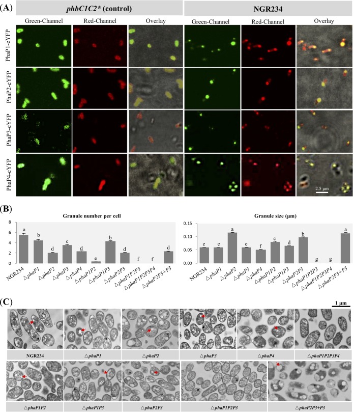
FIG 2.
Characteristics of PHB granules in free-living NGR234 and mutant strains lacking genes encoding phasins. (A) Subcellular colocalization of predicted phasins with PHB granules. PhaP1-eYFP, PhaP2-eYFP, PhaP3-eYFP, and PhaP4-eYFP fusions were constitutively expressed in free-living NGR234 and its phbC1C2 mutant (*, ΔphbC1C2 used for expressing PhaP4-eYFP). The phbC1C2 (or ΔphbC1C2) mutant served as a control with no PHB accumulation. Fluorescence microscopic images were generated after staining with Nile red in the red channel (indicating the localization of PHB granules) or without staining in the green channel (showing the localization of phasins). (B) Number of PHB granules within each bacterial cell and size of PHB granules. A total of 150 cells were scored for each strain (58 cells were scored for the ΔphaP1P2 mutant; these values were obtained under the same conditions as for Fig. 1, and the same values for NGR234 were used in this case). Values represent means ± SEMs. Different letters indicate significant difference (alpha = 0.05, Duncan’s test). (C) Transmission electronic microscopy pictures of bacterial cells at stationary phase. Red arrows indicate PHB granules. Bar, 1 μm.

