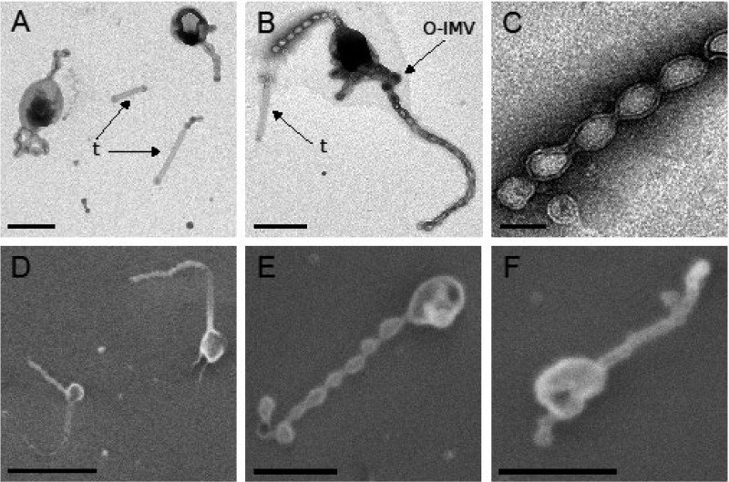
FIG 1.
Micrographs of strain Hel3_A1_48 cells in stationary growth phase. Membrane tubes (t) and thicker vesicles (assigned as O-IMVs) are indicated. Cells grown in HaHa_100V medium at 21°C to stationary phase were negatively stained with 1% uranyl acetate for transmission electron microscopy (TEM) (A to C). The cells were passively settled on a silica wafer, dehydrated by an ethanol series, and preserved using critical point drying for scanning electron microscopy (SEM) (D to F). Bar corresponds to 100 nm (C), 500 nm (A, B, E, and F), or 1 μm (D).

