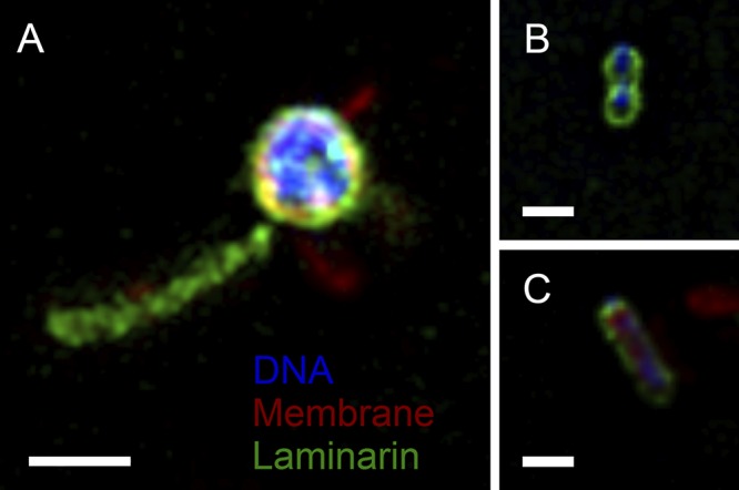
FIG 5.

Superresolution structured illumination microscopy image of strain Hel3_A1_48 in stationary growth phase reveals the label of laminarin (green color) on the outside cells (Nile red membrane stain, red color; DAPI DNA stain, blue color) and on appendages (A) and on dividing cells (B, C). Bars correspond to 1 μm.
