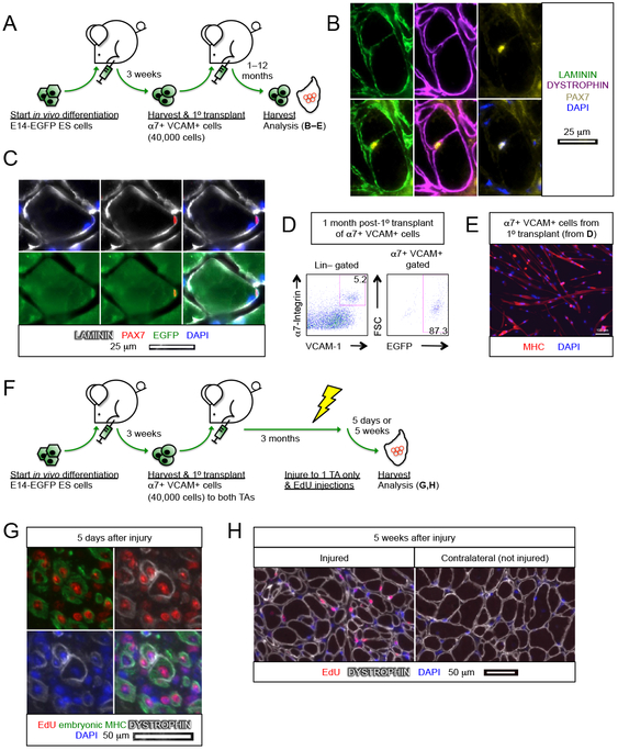
Figure 3. ES cell-derived myogenic progenitors reconstitute the muscle stem cell compartment.
(A) Schematic of evaluating the contribution of E14-EGFP ES cell-derived α7+ VCAM+ myogenic progenitors in the muscle stem cell compartment.
(B) PAX7+ cell associated with a DYSTROPHIN+ fiber under the basal lamina (n=6 biological replicates). Scale bar represents 25 μm.
(C) Donor derived PAX7+ EGFP+ muscle stem cell under the basal lamina (n=6 biological replicates). Scale bar represents 25 μm.
(D) FACS analysis of transplanted muscles revealed that the majority of α7+ VCAM+ muscle stem cells are also EGFP+, i.e., donor derived (n=7 biological replicates).
(E) Re-harvested α7+ VCAM+ EGFP+ cells (from C) differentiated into multi-nuclei MHC+ myotubes upon culture (n=7 biological replicates). Scale bar represents 100 μm.
(F) Schematic of evaluating the regenerative potency of transplanted E14-EGFP ES cell-derived α7+ VCAM+ myogenic progenitors upon subsequent injury.
(G,H) Transplanted α7+ VCAM+ cells regenerate upon subsequent injury. α7+ VCAM+ cells were transplanted into both left and right TA muscles and 3 months later only the left TA muscles were re-injured. EdU was administered 2–4 days after re-injury. Muscles were then harvested (G) 5 days or (H) 5 weeks after injury.
(G) The presence of EdU+ embryonic MHC+ fibers 5 days after re-injury indicates regeneration. Some EdU+ embryonic MHC+ fibers start to re-express DYSTROPHIN (n=4 biological replicates). Scale bar represents 50 μm.
(H) EdU+ DYSTROPHIN+ fibers in the injured muscle 5 weeks post-injury. Note that in the contralateral muscle that was not re-injured after the primary transplant, only EdU− DYSTROPHIN+ fibers were present (n=4 biological replicates). Scale bar represents 50 μm.
α7, α7-integrin. VCAM, VCAM-1. ES cells, embryonic stem cells. Lin, lineage cocktail comprising antibodies against CD45 (hematopoietic) and CD31 (endothelial). MHC, myosin heavy chain.

