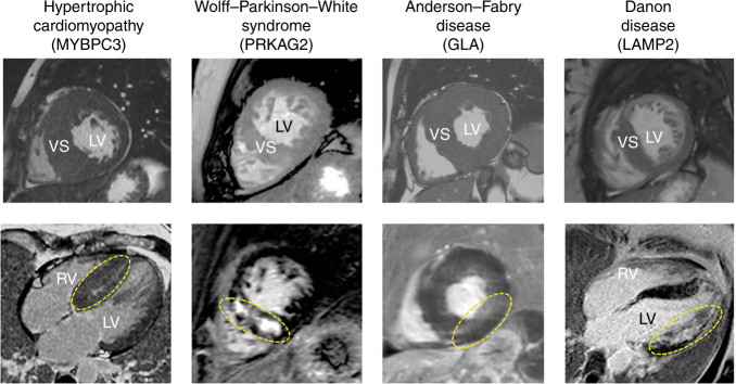
Fig. 3. Cardiac magnetic resonance images of example patients with sarcomeric hypertrophic cardiomyopathy and three of its mimics identified in our cohort.
Top: cine two-dimensional images in short-axis view using balanced turbo-field echo sequencing. Bottom: late gadolinium enhancement patterns (circled in yellow) in four-chamber (first and last panel) and short-axis view, using spoiled inversion recuperation turbo-field echo three-dimensional sequencing. LV left ventricle; RV right ventricle; VS ventricular septum

