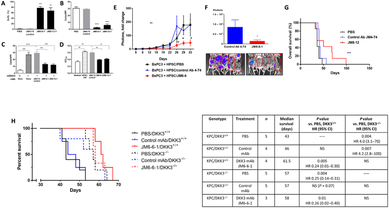
Fig. 5. DKK3-blocking Abs inhibit PSC and cancer cell activity, chemoresistance, and tumor progression, with improved survival.
HPSCs and BxPC3 cells were treated with DKK3 mAb clones JM6-6-1 and JM8-12-1 or isotype control mAb or PBS. (A and B) HPSC apoptosis (A) and migration (B) as measured by fluorescence-activated cell sorting (FACS) and Transwell migration assay at 48 hours. (C) BxPC3 migration in response to rhDKK3 (10 μg/ml) as measured by Transwell migration assay at 48 hours. (D) BxPC3 resistance to gemcitabine (100 μM) as measured by MTT proliferation assay at 6 days. HPSC-CM (10 μg/ml). (E) The orthotopic coinjection BxPC3 + HPSC model of PDAC was used to test the efficacy of DKK3 mAb clones JM6-6-1 or JM8-12-1 (5 mg/kg ip, once every 5 days). Overall tumor progression was measured every 3 to 4 days by IVIS imaging. (F) Metastatic tumors in the peritoneal cavity after removal of the primary pancreatic tumor are shown by IVIS imaging. (G) Kaplan-Meier survival curve showing mice treated with DKK3 mAb clone JM8–12 (red), control Ab (blue), or PBS (black). KPC mice (P48-Cre; Kras LSL-G12D;Trp53fl/fl) either with WT DKK3 (solid lines) or deficient in DKK3 (dashed lines) were treated with DKK3 mAb JM6-6-1 (5 mg/kg, ip, once every 5 days), PBS, or control mAb. Kaplan-Meier survival curve (H) is shown with hazard ratios (log-rank test). Data are means ± SEM [n = 7 mice per group in (E) and (F), 6 to 7 mice per group in (G), 3 to 5 mice per group in (H)]. *P < 0.05, **P < 0.01, ***P < 0.001, ****P < 0.0001 versus control Ab.

