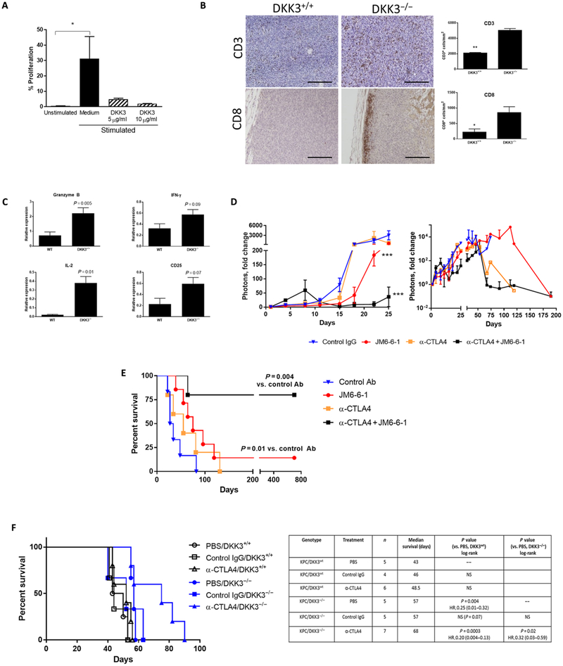
Fig. 6. DKK3 blockade is associated with increased tumor immune infiltrates and improves response to checkpoint inhibitor therapy.
(A) T cells were stimulated and treated with DKK3 (5 to 10 μg/ml), and proliferation was measured by carboxyfluorescein diacetate succinimidyl ester (CFSE) assay. In a syngeneic orthotopic model with luciferase-labeled KPC cells, (B) tumors were examined for CD3 and CD8 expression by IHC, and (C) additional markers of T cell activity were measured by qPCR. (D) Mice in the syngeneic orthotopic KPC-luciferase model were treated with control IgG, DKK3 mAb JM6-6-1, α-CTLA4, or the combination JM6-6-1 + α-CTLA4, and tumor growth was measured by IVIS imaging up to 190 days. Survival in this orthotopic implantation model is shown in (E). Using a GEMM (F), KPC/DKK3+/+ (black) or KPC/DKK3−/− (blue) mice were treated with α-CTLA4 or control IgG, and the Kaplan-Meier survival curve is shown. Data are means ± SEM [n = 5 mice per group in (B) and (C), 5 to 7 mice per group in (D) and (E), 4 to 7 mice per group in (F)]. *P < 0.05, **P < 0.01, ***P < 0.001. Scale bars, 200 μm.

