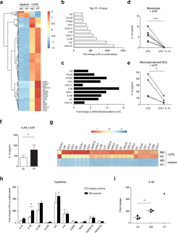
Figure 2. Dendritic cells with an IL10RA deficiency express high levels of IL-1β upon bacterial stimulation.
(a-c, g-i) Monocyte-derived dendritic cells from healthy controls (n=3), IBD patients (n=3) and an IL10RA-deficient patient (PT) were stimulated with LPS and high throughput RNA sequencing was performed after 6 hours of stimulation. (a) Heat map representing color-coded expression levels (FPKM values) of differentially expressed genes in IL10RA-deficient moDCs upon LPS stimulation. (b) Top 10 genes overexpressed in IL10RA-deficient moDCs upon LPS stimulation. Fold change values are calculated by dividing LPS-stimulated samples by unstimulated samples. (c) Comparison of the LPS-induced genes in (b) with expression levels in moDCs from healthy controls. (d, e) Monocytes and moDCs from adult healthy controls (n=4–6) were stimulated with LPS in the presence or absence of IL-10 for 20 h. Supernatants were assayed for IL-1β using an ELISA. To enhance IL-1β secretion, ATP was added during the last 15 minutes of stimulation. (f) Adult healthy controls (n=4) and IL10RA-deficient patient moDCs were stimulated with LPS for 20 h and ATP during the last 15 minutes of stimulation. Amount of IL-1β secreted by moDCs was determined by ELISA. (g) Heat map representing color-coded expression levels (FPKM values) of cytokine and chemokine genes in moDCs. (h, i) Cytokine mRNA expression in moDCs from (h) pediatric IBD patients and adult healthy controls and (i) adult healthy controls, pediatric IBD patients and IL10RA-deficient patient. Fold change values are calculated by dividing LPS-stimulated samples by unstimulated samples. Results are mean ± SD. *P<0.05, ***P<0.001 using one-way ANOVA (H, I) or unpaired Student’s t test (D, E, F). HC=healthy control, IBD=inflammatory bowel disease, PT=IL10RA-deficient patient.

