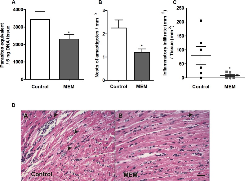
Fig 5. Tissue parasitic load, parasite density and inflammatory infiltrate in cardiac tissue.
(A) Measurement of parasitic load at 15 d.p.i. in tissues of BALB/c mice infected with 1x10³ forms of sanguine trypomastigotes and nontreated or treated with memantine (MEM) - 10 mg/kg per day—for ten consecutive days. The graph shows the number of parasites equivalent to 5 ng of tissue DNA. (B) Nests of amastigotes per mm2 in cardiac tissue at 15 d.p.i. BALB/c mice were infected with 1x10³ forms of blood trypomastigotes and treated with memantine (MEM) - 10 mg/kg per day—for ten consecutive days. The data are presented in number of nests per area of the analyzed section. (C) Cardiac tissue sections of 5 μm thick were obtained on 15 d.p.i., stained with H & E and analyzed by light microscopy. Areas of inflammatory infiltrates were quantified by an image analysis system. The sum of infiltrated areas on the six slides was calculated for each mouse. The final individual score was expressed in square micrometers of inflammatory infiltrates per square millimeter of area examined. * (p <0.05). (D) Histological view of the hearts of BALB/c mice infected with sanguine trypomastigotes and nontreated or treated with memantine (MEM). The experiments were repeated four times with 10 animals/group. The figure shows the presence of amastigotes nests (arrowheads). Scale bar represents 50 μm.

