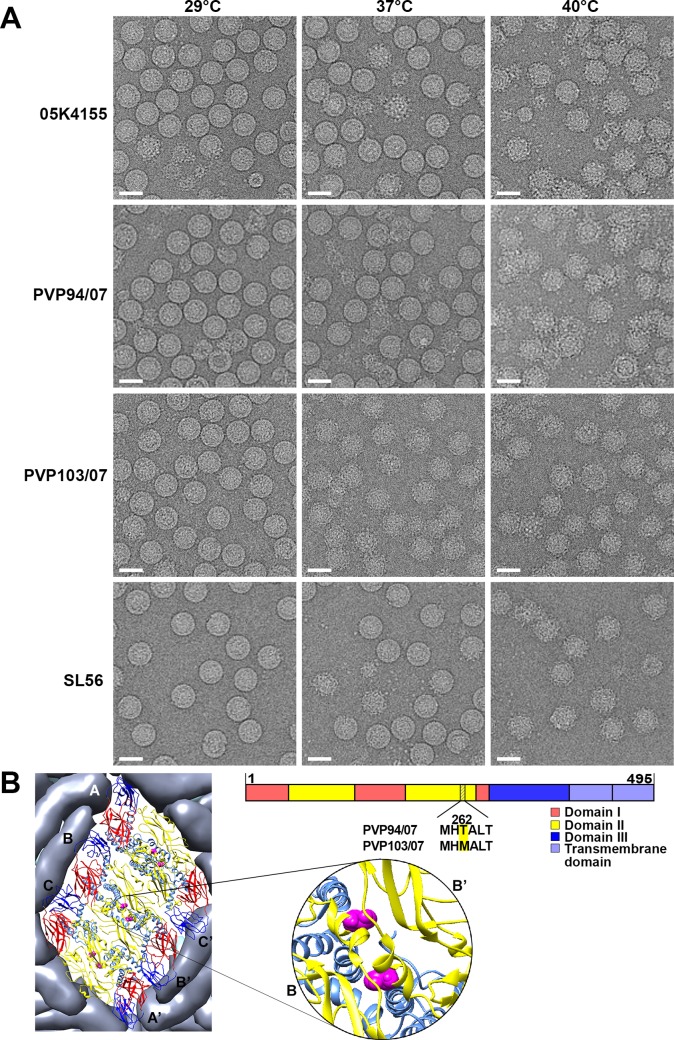
Fig 3. Morphology of DENV2 clinical strains at different temperatures.
(A) CryoEM micrographs of DENV2 clinical isolates (PVP94/07, PVP103/07, 05K4155 and SL56) at pH7.4 at 4°C, 37°C and 40°C. Scale bar is 50nm. (B) (Right) Sequence alignment of PVP94/07 and PVP103/07 showing only one residue difference at position 262 of E protein, and (left) its location on an E protein raft on the virus surface.

