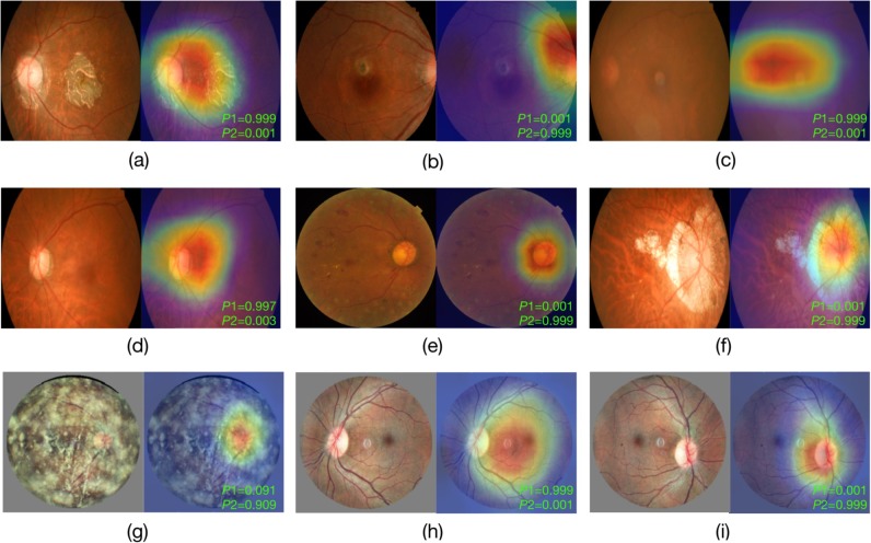
Fig 6. Visualization of the deep learning model on fundus images with different eye conditions.
The eye conditions from (a) to (g) are: (a) late dry age-related macular degeneration (AMD); (b) late wet AMD with disc tilt; (c) cataract; (d) glaucoma; (e) diabetic retinopathy; (f) high myopia; (g) vitreous opacity. (h) and (i) are two fundus images from both eyes of the same person. The bottom-right green text in each example indicates the corresponding probability scores outputted by the model (P1: left eye; P2: right eye).

