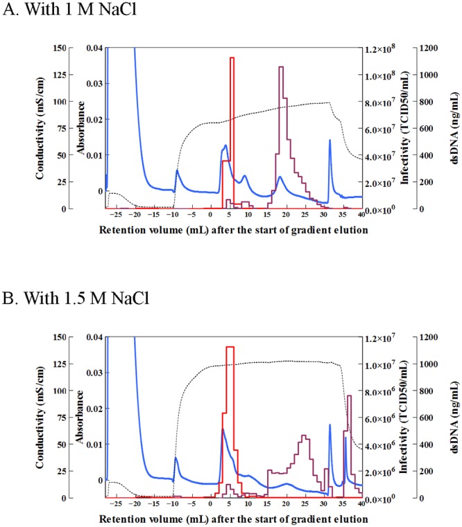
Fig 3. Chromatograms of Sabin type 2 virus-containing cell culture supernatant in the presence of NaCl.

Separation was carried out in the presence of (A) 1 M NaCl and (B) 1.5 M NaCl under the following conditions: column, ceramic hydroxyapatite (CHAp); sample (volume), cell culture supernatant containing Sabin type 2 virus (5 mL); buffer pH, 7.2; column wash, 10 mM sodium phosphate buffer (NaPB; 9 mL); equilibration, 10 mM NaPB with NaCl (14 mL); elution, linear gradient from 10 mM to 600 mM NaPB with NaCl for 30 mL; wash after separation, 600 mM NaPB (10 mL). Lines are the same as in Fig 2. TCID50, median tissue culture infectious dose.
