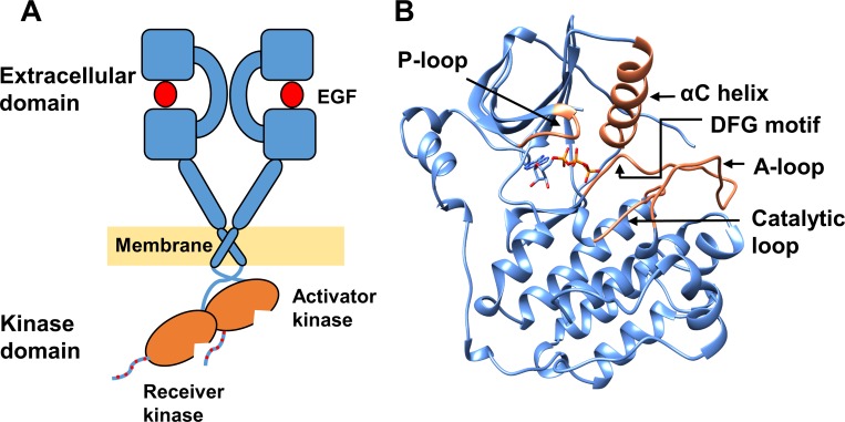
Fig 1. The EGFR structure.
(A) EGFR dimer–growth factor (red circle) -bound extracellular domains, transmembrane domains and asymmetrically dimerized intracellular kinase domains (orange) with phosphotyrosines at the C-terminal tail marked in red dots. (B) The kinase domain of EGFR (PDB ID 2ITX). Missing loops have been built using other EGFR structures. Bound ANP in the crystal structure was replaced by ATP (sticks). Key structural features are shown in orange.

