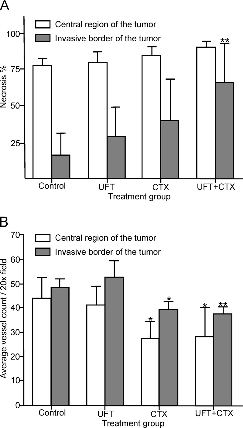
Fig 2. Tumor necrosis and vascular density are altered by the metronomic chemotherapy treatments.
(A) The percentage of tumor necrosis was assessed by H&E staining in sections of the central region and the invasive border of the untreated and treated tumors. A significant increase in tumor necrosis was observed in the invasive border sections of the UFT+CTX treated tumors (**p<0.01 vs. control); significance was analyzed by one-way ANOVA with Dunnett’s post-test. (B) Effect of the different treatments on vascular density as determined by double CD31/VEGR2 immunofluorescence labelling. The average vessel count of seven fields per tumor (x20 magnification), from the central region and the invasive border is presented. A significant reduction in vascular density was observed in the CTX (*p<0.05 vs. control) and the UFT+CTX (**p<0.01 vs. control) groups; significance was analyzed by one-way ANOVA with Dunnett’s post-test.

