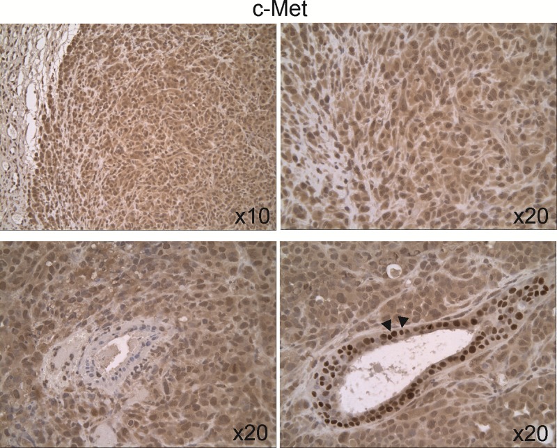
Fig 5. Expression and cellular distribution of c-Met in untreated 231/LM2-4 tumor xenografts.
c-Met staining was relatively homogenous throughout the tumor tissue, and was both cytoplasmic and nuclear. Hyperplastic acini showed cytoplasmic and nuclear staining, with strong staining in the nucleus (down-right arrows).

