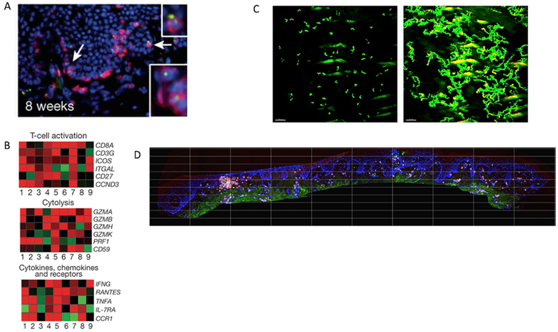
Figure 4. Immunosurveillance features of TRM include prolonged tissue residence, secretion of cytotoxic molecules, an activated transcriptional profile, intra-epithelial trafficking and self-organizational behavior.

(A) Immunohistochemical evidence of perforin granules (green) within multiple tissue-resident CD8+ T cells from human genital skin biopsies at a timepoint 8 weeks post lesion healing when HSV-2 antigen is not detected in tissue (reference 14) (B) Transcriptional evidence of an activated state in CD8+ T cells from human genital skin biopsies at a timepoint 8 weeks post healing when HSV-2 antigen is not detected in tissue. Gene expression is compared between CD8+ T cells detected at the dermal-epidermal junction (presumed TRM) versus control cells from the same individual with red and green indicating up and down regulated genes respectively (reference 14) (C) Intravital imaging evidence that TRM express dendritic arms and patrol extensively within but not beyond local micro-environment (reference 46). The left panel is an initial frame of a time-lapse movie in which the branching nature of TRM in murine flank skin is evident. The right panel is superimposed frames taken every 3 minutes over 7.5 hours showing that a majority of the local tissue region is patrolled. However, over more than a year, cells rarely migrate more than 5 mm from their starting location. (D) Immunohistochemistry evidence that TRM demonstrate self-organizational behavior in human genital skin with clustering of CD8+ T cells at the dermal epidermal junction. We determined that the ratio of CD8+ T cells to epithelial cells within the square regions follows an exponential slope on a rank order distribution, indicating a strict slope to CD8+ T cell structure in tissue.
