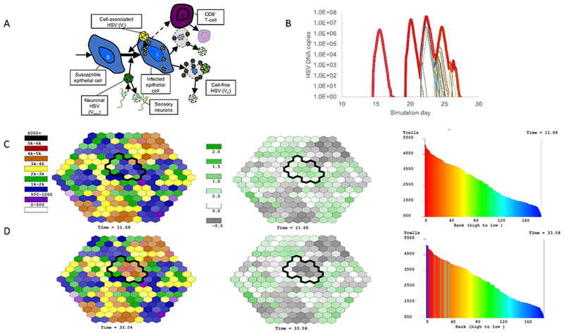
Figure 5. Mathematical model simulation of a prolonged HSV-2 shedding episode.

(A) Cartoon of mathematical model including susceptible cells, infected cells, cell-associated virus, cell-free virus and CD8+ T cells (TRM). These equations are simulated stochastically in 200 regions concurrently as shown in (C) & (D). (B) Simulated HSV-2 shedding episode notable for multiple peaks. The thick red line represents total viral load that would be detected with a clinical swab, while the thin lines represent viral load generated within single-microregions of infection. Simulated TRM structure and degree of protection (C) before and (D) following clearance of the shedding episode in (B). The left column represents the density of TRM within each micro-region before and after the episode. The circumscribed black regions had viral replication during the episode (thin lines in (B)). Regions which generated high viral loads transitioned from low to high TRM density as a large amount of local TRM proliferation was required to contain infection. The middle column demonstrates the log10 effective reproductive number (Re) of each region at the two timepoints. This number is inversely correlated with TRM density and denotes the potential for persistent cell-to-cell spread of virus prior to elimination. Green scale regions have Re>1 (log10 Re>0) indicating the need for TRM proliferation prior to elimination of all infected cells. Grey scale regions have Re<1 indicating no need for TRM proliferation prior to very rapid elimination of only a few infected cells. The right column plots the rank of each region according to TRM regional density. The shape of the distribution is stable before and after the episode, in keeping with the stable long-term détente established between HSV-2 and its host, despite dynamic reshuffling of individual region’s rank due to localized TRM proliferation events.
