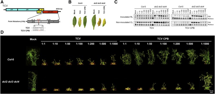
Figure 1.
TCV infection-caused disease symptoms in Arabidopsis. A, Schematic representation of the TCV genome showing the CPB point mutation on the CP (P38). Top diagram shows the genomic RNA of TCV. The bottom diagram shows the CP (P38) region of single-amino acid mutant TCV CPB. The single amino acid change in TCV CPB mutant is marked beneath the bottom diagram. The bottom diagram represents the full-length CP, with the sizes and the relative positions of the five structural domains: N, N terminal; R, RNA-binding domain; A, arm domain; S, surface domain; hinge; P, protruding domain; C, C terminal. B, TCV infection-caused chlorosis in noninoculated cauline leaves. Left: wild-type control (Col-0); right: hypersusceptible control (dcl2 dcl3 dcl4). Photographs were taken at 14 dpi. C, Local and systemic accumulation of CP (P38) caused by TCV (left) and TCV CPB (right) infection was assayed by immunoblotting. P38 was detected using anti-P38 antibody. Rubisco protein was detected by anti-Rubisco antibody as a control. The virion inoculum was in different dilutions. RL, rosette leaf; CL, cauline leaf. RL samples were collected at 7 dpi; CL samples were collected at 14 dpi. D, TCV infection-caused stunt bolt phenotype in Arabidopsis. Upper: wild-type control (Col-0); bottom: hypersusceptible control (dcl2 dcl3 dcl4). The virion inoculum was in different dilutions. Photographs were taken individually at 14 dpi and digitally extracted and aligned for comparison.

