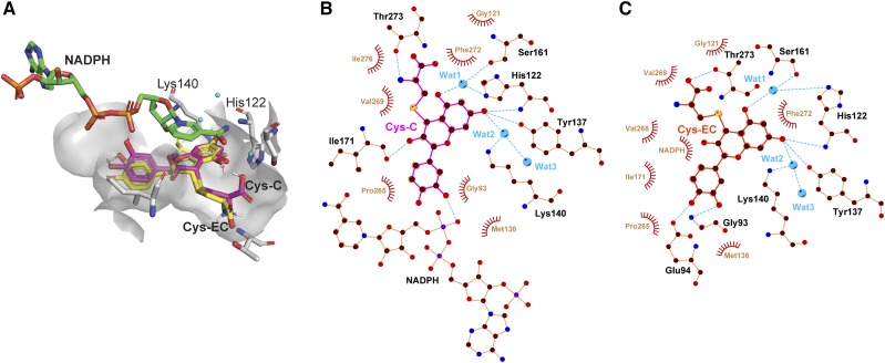
Figure 5.
Molecular docking analysis of Cys-C and Cys-EC in the VvLAR1 active site. A, Superimposed 3D structures of Cys-C and Cys-EC docked with VvLAR1 (PDB ID: 3I52) active site. The ligands NADPH, Cys-C, and Cys-EC and catalytic residues His-122 and Lys-140 are shown as bond models, and the three water molecules are shown as blue spheres. B and C, ligand-protein interaction diagrams of VvLAR1 with Cys-C and Cys-EC as substrates. Water molecules (Wat) are shown as blue balls. Hydrogen bonds are shown as blue dotted lines, while the spoked arcs represent protein residues or NADPH making nonbonded contacts with Cys-C and Cys-EC.

