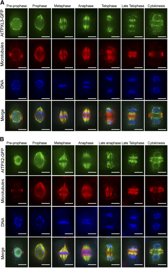
Figure 4.
TPX2 and TPXL3 localize differentially in dividing Arabidopsis root cells. A and B, TPXL3-GFP (A) and TPX2-GFP (B) localization in dividing Arabidopsis root cells obtained via immunofluorescence with antibodies against GFP (green), MTs (red), and DNA (blue) in cells from prophase to telophase. Before NEBD, TPXL3 largely accumulates at the NE, while TPX2 is mostly nuclear. Both proteins are not detectable at the PPB. After NEBD, both TPX2 and TPXL3 decorate the two “polar caps” of the prophase spindle as well as spindle MTs and the shortening K-fibers at anaphase. In late anaphase and telophase, when midzone MTs develop into the two mirrored sets of the early phragmoplast array, TPX2 association with the phragmoplast-forming MTs is much more pronounced than that of TPXL3, which remains heavily enriched at the former spindles poles and subsequently localizes to the reformed NE with a strong bias toward the part facing the reforming daughter nuclei. Scale bars = 5 μm.

