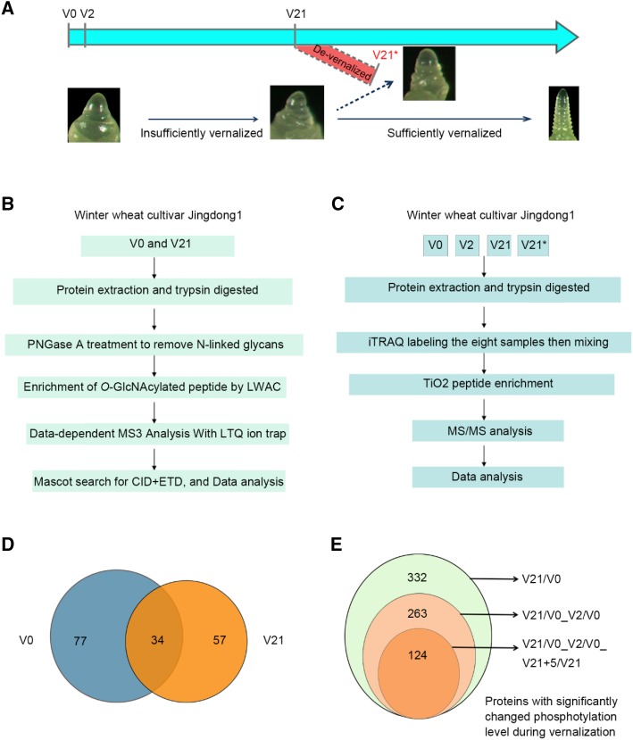
Figure 2.
Experiments design to enrich and identify proteins with O-GlcNAcylation or phosphorylation at different stage of vernalization and overview of identified proteins. A, Diagram of tissue sampling at different time point during vernalization and the corresponding developmental stages of shoot apex. B and C, Strategies used for isolation, enrichment, and identification of O-GlcNAcylated peptide/protein (B) and phosphorylated peptide/protein with two biologic replications (C). D, Venn diagram showing general and unique O-GlcNAcylated proteins identified at V0 and V21. E, Venn diagram showing alternatively changed phosphorylated proteins identified in response to vernalization. V21/V0, protein of SCPL between V21 and V0; V21/V0_V2/V0, SCPL protein between V21 and V0 deduct that of V2 and V0; V21/V0_V2/V0_V21+5/V0, SCPL protein between V21 and V0 subtract that of V2/V0 and V21+5/V0. V0: no cold exposure; V2: vernalization for 2 d; V21: vernalization for 21 d; and V21*: vernalization for 21 d followed by high temperature (35°C) growth for 5 d (V21+5). CID, collisional induced dissociation; ETD, electron transfer dissociation.

