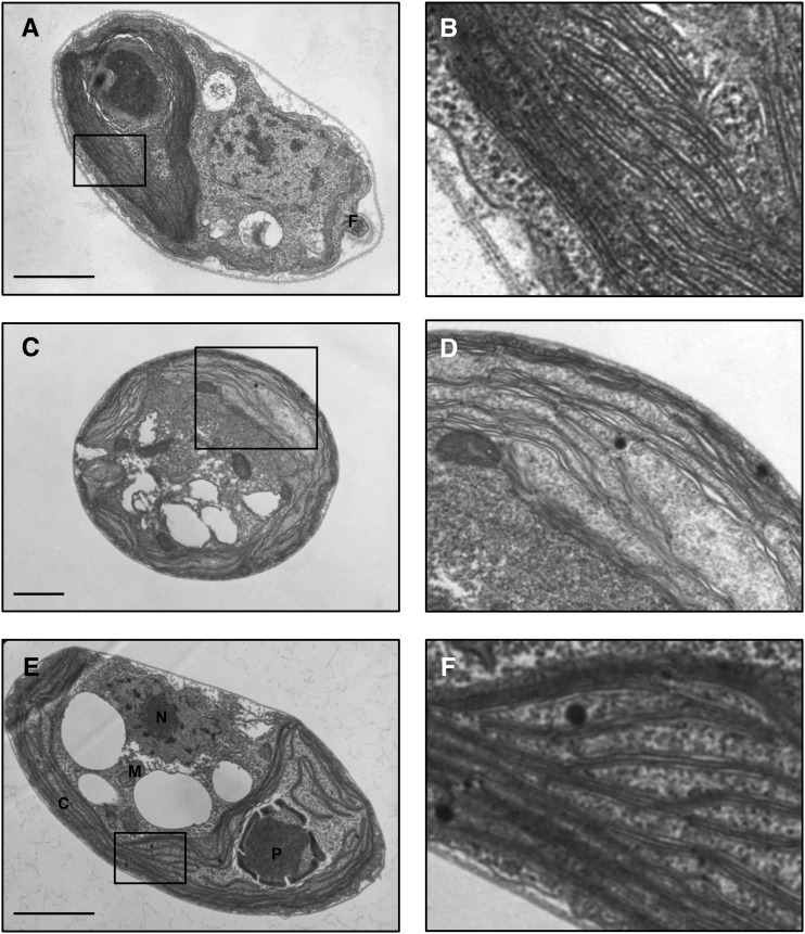
Figure 11.
Typical electron micrographs of C. reinhardtii and Clamydomonas sp. UWO241 are shown. The images in the right column are magnifications of the boxed areas in the left column for midlog phase cells of C. reinhardtii grown in BBM at 23°C (A and B), UWO241 grown in HS (700 mm NaCl) at 5°C (C and D), and UWO241 grown in LS (70 mm NaCl) at 5°C (E and F). Scale bars = 1.0 µm. C, chloroplast; N, nucleus; M, mitochondrion; P, pyrenoid.

